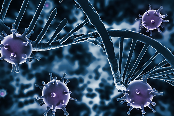
Parkinson’s disease (PD) is a progressive neurodegenerative disorder affecting dopamine-producing neurons in the substantia nigra. At its core, PD is driven by the accumulation and spread of α-synuclein (αSyn) into toxic aggregates known as Lewy bodies. Yet, the mechanisms governing this spread remain poorly understood, and no approved treatment exists to slow disease progression.
To address this, researchers at the Van Andel Institute and Michigan State University set out to map the molecular landscape of PD neurons across disease stages. Our analytical team at VUGENE supported this effort through advanced statistical modeling and pathway-level interpretation, enabling the identification of novel mechanisms underlying disease progression.
Deep Molecular Characterization
RNA sequencing of neuronal nuclei isolated from the prefrontal cortex of 105 individuals (spanning early-stage PD, late-stage PD, and healthy controls) identified 6,481 differentially expressed genes. By focusing on sorted neurons rather than bulk tissue, the analysis captures disease-relevant changes with greater precision than prior studies.
Ciliogenesis as an Unexpected Player
The most striking finding was a consistent upregulation of primary cilia gene networks, structures that act as cellular antennae for signal detection, present in both early- and late-stage PD. This pattern, spanning ciliogenesis, axoneme structure, and SHH signaling, suggests a compensatory neuronal response that persists across disease progression.
From Observation to Mechanism
Boosting ciliogenesis in vitro using the SHH-pathway activator purmorphamine produced a dose-dependent reduction in αSyn pathology. Deletion of TET2, an epigenetic regulator increasingly linked to PD, replicated this effect in vivo, reducing αSyn spread, enhancing cilia-related gene expression, and protecting dopamine neurons from degeneration.
A New Therapeutic Direction
Together, these findings connect epigenetic regulation through TET2 with ciliary biology and αSyn pathology. One likely mechanism is autophagy: enhanced ciliogenesis may improve the neuron’s ability to clear toxic protein aggregates. Because ciliary alterations are observed across disease stages, they may offer stage-specific windows for intervention.
The Bioinformatics Behind the Breakthrough
To identify differentially expressed genes, we applied robust linear regression via the limma statistical framework, adjusting for age, sex, brain hemisphere, postmortem interval, and sample origin. Pathway enrichment analysis including GSEA and Enrichment Map-based network clustering translated thousands of gene-level changes into a structured biological map of PD progression and produced the publication-ready visualizations featured in the paper.
Why Does This Matter?
This study demonstrates that ciliogenesis is not a bystander in Parkinson’s disease. It is an active, potentially protective process that neurons deploy in response to αSyn pathology. By identifying TET2 as an upstream epigenetic regulator of this response, the work opens a new avenue for therapeutic development. Future research will determine whether enhancing ciliogenesis pharmacologically can meaningfully slow disease progression in patients.
Emmanuel Quansah, Naman Vatsa, Elizabeth Ensink, Jaycie Brown, Tyce Cave, Miguel Aguileta, Emily Schulz, Allison Lindquist, Carla Gilliland, Jennifer A. Steiner, Martha L. Escobar Galvis, Milda Milčiūtė, Michael X. Henderson, Patrik Brundin, Lena Brundin, Lee L. Marshall & Juozas Gordevičius (2025). Tet2 loss and enhanced ciliogenesis suppress α-synuclein pathology. Acta Neuropathologica Communications, vol 13 (1).
Read full article: Link
Cover image credits: R Photography / Adobe Stock